Search for echo echocardiogram. check all results web now!. M-mode echocardiographic measurements. m-mode echocardiography has good temporal resolution. hence measurements of the left ventricle are often taken with m-mode. end diastolic and end systolic volumes are estimated from these measurements. the stroke volume and ejection fraction are also calculated from these measurements. Two-dimensional and m-mode echocardiographic measurements of cardiac dimensions in healthy standardbred trotters acta vet hung. 2002;50(3):273-82. doi: 10. 1556/avet. 50. 2002. 3. 3.
M-mode echocardiography, however, does retain a role in functional echocardiography and, due to its superior temporal and spatial resolution, is most helpful when used for the timing of rapid cardiac motion and precise linear measurements of cardiac dimensions. M-mode echocardiography has been used for measurements of lv size, mass, and loading conditions (end-systolic meridional and circumferential wall stress), and m-mode echocardiography measurements myocardial function (fractional and mid-wall shortening and velocity of circumferential fiber shortening peak).
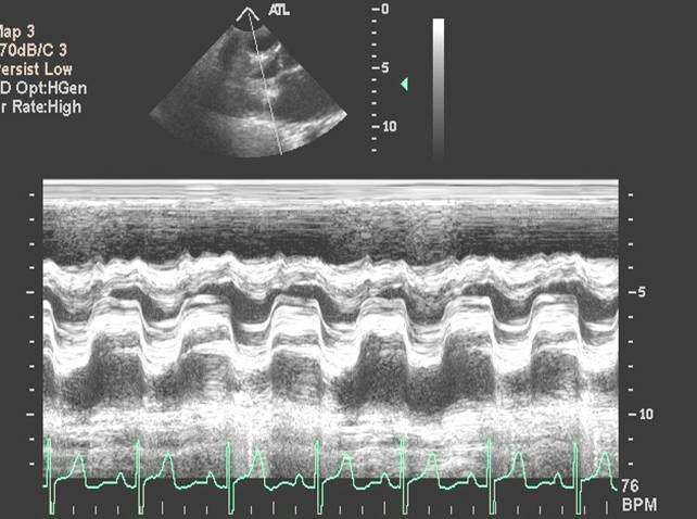
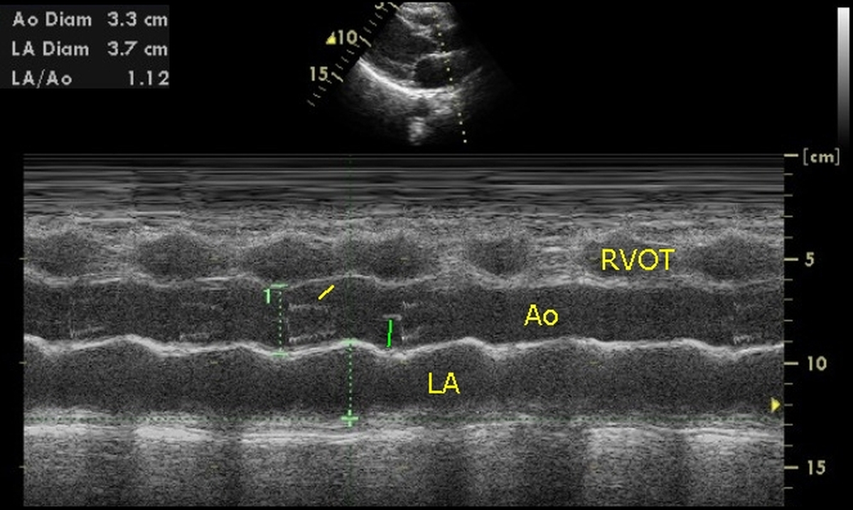
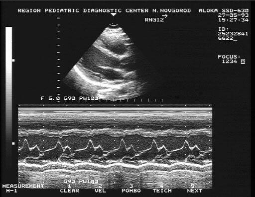
Direct digital measurements, while better two dimensional resolution avords more accurate placement of the m mode beam. our aim in this study was to re-evaluate the limits of echocardiographic measurements of 8 7. 5 7 6. 5 6 5. 5 5 4. 5 4 3. 5 3 2. 5 2 1. 5 1 0. 5 0 right ventricular anterior wall thickness at end diastole (mm) 1. 9. M-mode imaging the m-mode echo, which provides a 1d view, is used for fine measurements. temporal and spatial resolutions are higher because the focus is on only one of the lines from the 2d trace (see figure 2). figure 2.
Using m-mode in echocardiography displays the movement of the myocardium allowing for accurate and real-time measurements of wall thickness, internal diameter, . Recommendations regarding quantitation in m-mode echocardiography: results of a survey of m-mode echocardiography measurements echocardiographic measurements. d j sahn;; a demaria;; j kisslo; .

Diastolic velocity in long-axis direction obtained by m-mode echocardiography and by tissue doppler in the as-sessment of right ventricular diastolic function. clin physiol funct imaging. 2005;25(3):178-182. iv. loiske k, waldenborg m, fröbert o, rask p, emilsson k. left and right ventricular systolic long-axis function and. M-mode echocardiography m-mode was previously the dominating modality in echocardiography. although it has now largely been replaced by 2d echocardiography, it is still used in clinical practice. m-mode provides a one-dimensional view of all reflectors (i. e structures reflecting ultrasound waves) along one ultrasound line.
M-mode m-mode echocardiography measurements echocardiography. m-mode was previously the dominating modality in echocardiography. although it has now largely been replaced by 2d echocardiography, it is still used in clinical practice. m-mode provides a one-dimensional view of all reflectors (i. e structures reflecting ultrasound waves) along one ultrasound line.
Normal Values Of M Mode Echocardiographic Measurements Of
Compared with m-mode echocardiography, 2d echocardiography offers improved spatial orientation because the left ventricle can be sampled in real time at multiple levels. accordingly, 2d echocardiography may be used to quantify the lv mass and chamber volumes of abnormally shaped ventricles, and because measurements of lv length are possible. Both bidimensional and m-mode echocardiography are useful in small rodent models, particularly for the serial assessment of lv chamber size, mass, and systolic function. 9 in addition, pulsed-wave doppler (pwd) echocardiography has been used in these models for the in vivo assessment of diastolic function 10,11 and global lv performance. 12,13 however, a reference range of echocardiographic values reflecting normal lv function in hamsters is still lacking. M mode echocardiography was the first tte application used to assess la size. the la internal dimensions should be measured at lv end systole when the la .
M-mode at the mitral valveamplitude descriptionnormalvalueepss measure e point to septalseparation< 5 mmd-e measures the maximumexcusion of the mitral . Ography (2de)-guided m-mode approach, although linear measurecalculate lv mass from m-mode echocardiography, 2de, and 3de. (table 5). all measurements .
M-mode echocardiography and 2d cardiac measurements.
M-mode echocardiographic measurements m-mode echocardiography has good temporal resolution. hence measurements of the left ventricle are often taken with m-mode. end diastolic and end systolic volumes are estimated from these measurements. the stroke volume and ejection fraction are also calculated from these measurements. Ican society of echocardiography standards documents, except for m-mode measurements, which have limited value. the superior temporal resolution of m-mode .

Download scientific diagram m-mode echocardiography measurement of the septum, posterior wall, lv end-systolic and end-diastolic diameter. M-mode imaging. the m-mode echo, which provides a 1d view, is used for fine measurements. temporal and spatial resolutions are higher because the focus is on only one of the lines from the 2d trace (see figure 2). Objective to obtain normal m mode (one dimensional) echocardiographic values in a substantial sample of normal infants and children. design data were obtained over three years from a single centre in central europe. patients 2036 healthy infants and children aged one day to 18 years. methods in line with recommendations for standardising measurements from m mode echocardiograms, and using. Objective to obtain normal m mode (one dimensional) echocardiographic values in a substantial sample of normal infants and children. design data were obtained over three years from a single centre in central europe. patients 2036 healthy infants and children aged one day to 18 years. methods in line with recommendations for standardising measurements from m mode echocardiograms, and using.
M-mode echocardiography the original description of m-mode echocardiography in 1953 by dr. inge edler and physicist hellmuth hertz marked the beginning of a . Find cardiac echocardiography. get high level of information!. While 2d echocardiography is essentially a “picture” of the heart, an m-mode echocardiogram is a “diagram” that shows how the positions of its structures change during the cardiac cycle. m-mode recordings allow in-vivo noninvasive measurement of cardiac dimensions and motion patterns of its structures.
Understanding The Echocardiogram Cardiology Explained
Society of echocardiography. j am soc echocardiogr 2010;23:685-713. 3. lopez l, colan sd, frommelt pc, ensing gj, kendall k, younoszai ak, lai ww, geva t. recommendations for quantification methods during the performance of a pediatric echocardiogram: a report from the pediatric measurements writing group of the american society of echocardiography. Search what is echocardiogram test. compare results on allproductsweb.

to a m-mode and two-dimensional echocardiogram with color doppler the analyses were restricted to healthy participants echocardiographic measurements Key words: canine; echocardiography; heart; reference range. m-mode echocardiography is commonly used to m-mode echocardiography measurements measure linear cardiac dimensions of cardiac chambers .


0 Response to "M-mode Echocardiography Measurements"
Post a Comment